Home
/ Tendon Diagram : Flexor Tendon Injuries Orthoinfo Aaos - However, this condition also involves the failure of associated ligaments and joints on the medial (inner) side of the foot and ankle.
Tendon Diagram : Flexor Tendon Injuries Orthoinfo Aaos - However, this condition also involves the failure of associated ligaments and joints on the medial (inner) side of the foot and ankle.
Tendon Diagram : Flexor Tendon Injuries Orthoinfo Aaos - However, this condition also involves the failure of associated ligaments and joints on the medial (inner) side of the foot and ankle.. The two peroneal tendons in the foot run side by side behind the outer ankle bone. When the biceps contracts, it pulls the forearm up and rotates it outward. In man, some 10 to 20 muscle fibres are connected to one tendon organ. Tendon reflexes, also referred to as myotatic reflexes, are variably affected by lower motor neuron lesions. The achilles tendon is a band of tissue that connects a muscle to a bone.
If the complete motor neuron innervation to a muscle is lost, the efferent limb of a tendon reflex will fail. The long head of biceps tendon is usually located inferiorly in the bicipital groove held there by the biceps pulley (the stabilization role of the transverse humeral ligament is controversial) 3. If you would like to learn all the parts of the foot structure, you have come to the right place. Understanding the structure of the foot is best done by looking at a foot diagram where the anatomy has been labeled. In many cases, torn tendons begin by fraying.
Patellar Tendonitis Jumper S Knee Johns Hopkins Medicine from www.hopkinsmedicine.org Hypoactive or absent reflexes are commonly observed in association with lower motor neuron lesions. One peroneal tendon attaches to the outer part of the midfoot, while the other tendon runs under the foot and attaches near the inside of the arch. If you would like to learn all the parts of the foot structure, you have come to the right place. It runs down the back of the lower leg and connects the calf muscle to the heel bone. The long head of biceps tendon is usually located inferiorly in the bicipital groove held there by the biceps pulley (the stabilization role of the transverse humeral ligament is controversial) 3. If the complete motor neuron innervation to a muscle is lost, the efferent limb of a tendon reflex will fail. In man, some 10 to 20 muscle fibres are connected to one tendon organ. However, this condition also involves the failure of associated ligaments and joints on the medial (inner) side of the foot and ankle.
The two peroneal tendons in the foot run side by side behind the outer ankle bone.
The long head of biceps tendon is usually located inferiorly in the bicipital groove held there by the biceps pulley (the stabilization role of the transverse humeral ligament is controversial) 3. In this area it is confined to a tight tunnel. Biceps tendon tears can be either partial or complete. Dislocation of the long head of biceps tendon is a common shoulder injury. The achilles tendon is a band of tissue that connects a muscle to a bone. However, this condition also involves the failure of associated ligaments and joints on the medial (inner) side of the foot and ankle. Understanding the structure of the foot is best done by looking at a foot diagram where the anatomy has been labeled. A typical tendon organ in limb muscles has an ending of about 0.5 mm in length. In many cases, torn tendons begin by fraying. A tendon is a band of tissue that connects a muscle to a bone. A complete tear will split the tendon into two pieces. The two peroneal tendons in the foot run side by side behind the outer ankle bone. As the damage progresses, the tendon can completely tear, sometimes when lifting a heavy object.
Hypoactive or absent reflexes are commonly observed in association with lower motor neuron lesions. The tendon runs around a bony prominence on the back of the wrist called lister's tubercle. Achilles tendon disorders what is the achilles tendon? The tendon that attaches the biceps muscle to the forearm bones (radius and ulna) is called the distal biceps tendon. This tendon is vulnerable to rupture in the tunnel at the wrist.
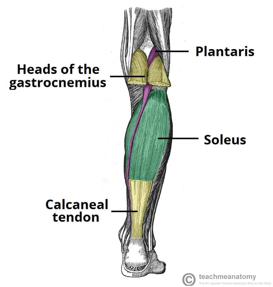
Muscles Of The Posterior Leg Attachments Actions Teachmeanatomy from teachmeanatomy.info It runs down the back of the lower leg and connects the calf muscle to the heel bone. If the complete motor neuron innervation to a muscle is lost, the efferent limb of a tendon reflex will fail. This tendon is vulnerable to rupture in the tunnel at the wrist. A complete tear will split the tendon into two pieces. Also called the heel cord, the achilles tendon facilitates walking by helping to raise the heel off the ground. The tendon runs around a bony prominence on the back of the wrist called lister's tubercle. Jul 05, 2018 · the foot diagram has a complex structure made up of bones, ligaments, muscles, and tendons. If you would like to learn all the parts of the foot structure, you have come to the right place.
In this area it is confined to a tight tunnel.
Dislocation of the long head of biceps tendon is a common shoulder injury. This tendon is vulnerable to rupture in the tunnel at the wrist. The achilles tendon is a band of tissue that connects a muscle to a bone. Achilles tendon disorders what is the achilles tendon? In this area it is confined to a tight tunnel. The tendon that attaches the biceps muscle to the forearm bones (radius and ulna) is called the distal biceps tendon. Tendon reflexes, also referred to as myotatic reflexes, are variably affected by lower motor neuron lesions. (see diagram) why does it rupture? Also called the heel cord, the achilles tendon facilitates walking by helping to raise the heel off the ground. As the damage progresses, the tendon can completely tear, sometimes when lifting a heavy object. Many tears do not completely sever the tendon. Biceps tendon tears can be either partial or complete. The tendon runs around a bony prominence on the back of the wrist called lister's tubercle.
This tendon is vulnerable to rupture in the tunnel at the wrist. The tendon runs around a bony prominence on the back of the wrist called lister's tubercle. In this area it is confined to a tight tunnel. Biceps tendon tears can be either partial or complete. The long head of biceps tendon is usually located inferiorly in the bicipital groove held there by the biceps pulley (the stabilization role of the transverse humeral ligament is controversial) 3.
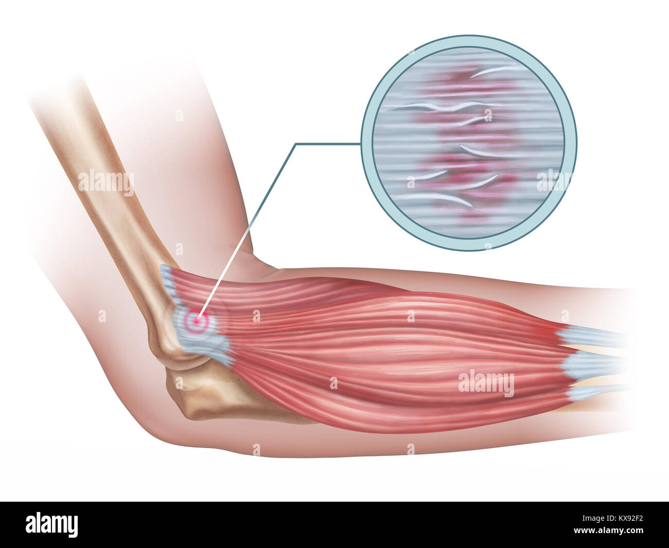
Tennis Elbow Diagram Showing A Detail Of The Damaged Tendon Tissue Digital Illustration Stock Photo Alamy from c8.alamy.com This tendon straightens the end joint of the thumb and also helps pull the thumb in towards the index finger. In many cases, torn tendons begin by fraying. A complete tear will split the tendon into two pieces. The tendon runs around a bony prominence on the back of the wrist called lister's tubercle. Jul 05, 2018 · the foot diagram has a complex structure made up of bones, ligaments, muscles, and tendons. When the biceps contracts, it pulls the forearm up and rotates it outward. Hypoactive or absent reflexes are commonly observed in association with lower motor neuron lesions. A tendon is a band of tissue that connects a muscle to a bone.
In this area it is confined to a tight tunnel.
A complete tear will split the tendon into two pieces. A typical tendon organ in limb muscles has an ending of about 0.5 mm in length. Biceps tendon tears can be either partial or complete. Understanding the structure of the foot is best done by looking at a foot diagram where the anatomy has been labeled. This tendon is vulnerable to rupture in the tunnel at the wrist. The two peroneal tendons in the foot run side by side behind the outer ankle bone. (see diagram) why does it rupture? In many cases, torn tendons begin by fraying. If the complete motor neuron innervation to a muscle is lost, the efferent limb of a tendon reflex will fail. It runs down the back of the lower leg and connects the calf muscle to the heel bone. This tendon straightens the end joint of the thumb and also helps pull the thumb in towards the index finger. One peroneal tendon attaches to the outer part of the midfoot, while the other tendon runs under the foot and attaches near the inside of the arch. In man, some 10 to 20 muscle fibres are connected to one tendon organ.